Excitable Systems
The Hodgkin–Huxley and the Mitchell–Shaeffer models both are part of a class of excitable systems. The excitability arises from a threshold–like behavior that can drive the system into an excited state from which it tends to return after a characteristic amount of time. The two models also have a characteristic refractory period after the excited state which prevents the system from being re–excited for some duration of time.
The pioneering work of Hodgkin and Huxley, for which they received the Nobel Prize, first established the basic nonlinear system of equations for describing the transmembrane potential (voltage) across a small patch of the cell membrane in the giant axon of the squid. More generally, this marked the introduction of a general formalism for describing the dynamics of the membrane potential of any excitable cell that is governed by the various ionic currents crossing the membrane. Such models have been extended beyond neurons to cardiac cells, myocytes, electrocytes, endocrine cells, pancreatic beta cells, etc., and are sometimes referred to as conductance–based models or Hodgkin–Huxley–like models.
Spatial Extension and Reaction–Diffusion Equations
In the case of cardiac tissue, cardiac cells interact with their nearest neighboring cells through electrical connections called gap junctions which can be described mathematically in a way that is similar to resistors in electronics. Such a set of connections within a two–dimensional grid, taken in the limit of a spatial continuuum, effectively produces a Laplacian operator which is used in the modeling of diffusion processes. However, each cell additionally interacts with the extracellular medium surrounding it through a variety of ion channels where (i) the flow of ionic current across the channel is governed by electrical and chemical gradients across each channel and also (ii) the channels are opened and closed, or “gated”, in highly nonlinear ways, e.g., by voltage, calcium, ligands, neurotransmitters, etc.
Therefore, the dynamics of the spatially–extended systems is governed by two major effects: ‘diffusion’ across space due to cell–to–cell coupling, and excitability due to the intrinsic, nonlinear ionic currents that move across the membranes of the cardiac cells. This ability to become excited and return to an unexcited state shares many similarities with chemical reactions and these models can be included amongst the class of reaction–diffusion equations which arose historically in the context of chemical reactions and are capable of rich, spatiotemporal dynamic behavior, e.g., spiral waves.
Information for the movies
· Click image to download movie
· Movies are mpeg–4 basic (.mp4)
|
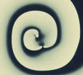
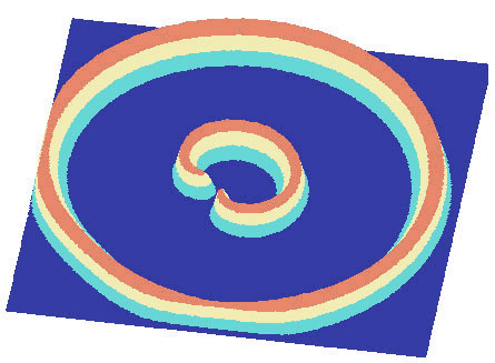
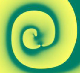
Spiral Wave Break–Up and Defibrillation
Simulations were performed on a spatially–extended version of the (modified) two–variable Mitchell–Shaeffer
model for cardiac tissue. Instabilities inherent in this model prevent a self–sustained spiral. Instead the sprial breaks up, continually generating new spiral
doublets which rapidly usurp the domain. Though it is not clear whether break–up of a spiral wave is the cause of fribrillation in
heart tissue, it is thought that the field of doublets, generated in possibly a different way, is representative of fribrillation.
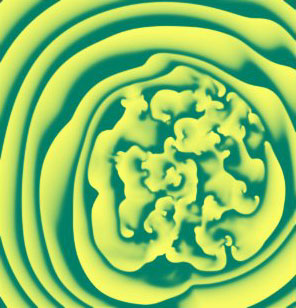
Defibrillation is then the extinguishing of this self–sustained activity, which prevents the normal functioning of the heart, and is accomplished by
the application of electrical currents to the surface of the body (the paddles). How such external currents manifest themselves within the heart to induce
defibrillation remains a heated debate ... i.e., a great mystery to be solved. The tail end of the movies demonstrates one approach to
defibrillation, proposed by James P. Keener, which asserts that small–scale inhomogeneities in the tissue set up small dipole currents whose size, relative to the spiral doublets’ cores, is an important factor to explain the experimental data.
|
|